From Atlas Obscura:
If “Harriet” could hear, she might pick up the sound of ping-pong balls skittering across a table. If she could smell, she might detect a range of lunches being reheated in a nearby microwave. If her eyes could see, she might let them wander a busted Pac-Man machine, a TV, and a campus bookstore, decorated with a swooping, celebratory paper chain, like an elementary-school version of DNA’s double helix. She might even catch a glimpse of herself in a camera lens or an observer’s glassy eyeballs. People often stop to stare.
On a sweaty Saturday, before social distancing was the law of the land, a group of visitors gathered at Drexel University’s medical campus in Northwest Philadelphia to meet “Harriet.” The preamble to this encounter was a display case holding several unusual and meticulously prepared medical specimens, long used as teaching tools. Like “Harriet,” each had been created in the late 19th century by a star anatomist, Rufus Weaver. Now, behind glass, between the cadaver lab and a bookstore, a segment of intestine and a piece of a spinal cord sit in stillness. A dissected eyeball floats ethereally in century-old liquid, its separated parts looking like a tiny jellyfish, a bit of brittle plastic, a mushroom cap.
The visitors shuffled through the door and into the otherwise empty student center. They huddled on the low-pile carpet, nondescript in the style of a suburban office park, and peered at more of Weaver’s dissection work, which occupied a glass-fronted case. They surveyed a sinewy hand, ropey and purplish. Two skulls and necks. Then, “Harriet.”
Reactions rippled.
“Oh.”
“Oh, wow.”
Quietly, “Poor Harriet.”
. . . .
“I’ve been meaning to find her,” said Malaya Fletcher, an epidemiologist in Washington, D.C., specializing in infectious disease. Fletcher remembered learning about the dissection in her high school biology class, and the story had stuck with her. “It’s just awesome,” she said. “You almost don’t believe it’s real.” The group crowded in close, lofting their cell phones above each other’s heads. They bobbed and weaved their raised hands, trying to take pictures without capturing their own flushed faces reflected in the glass.
“Harriet” is a network of fibers fastened to a black board in a case pushed up against a wall. At the top, there appears to be a brain, plump and brown, and a pair of eyes. Scan your own eyes down and you’ll encounter an intricate system of skinny, brittle cords, pulled taut and painted startlingly, artificially white. The outline is recognizably human—there’s the impression of hands and feet, the hint of a pelvis, the suggestion of a rib cage—but it is slightly fantastical, too. The way the cords loop at the hands and feet, it almost appears as if the figure has fins. Elsewhere, the fibers look shaggy, like chewed wire, as if electricity is shooting from the margins of the body.
This is a human medical specimen, in the spirit of an articulated skeleton. But unlike that familiar sight, it represents the nervous system, a part of the body’s machinery that most people have trouble even imagining. Some who stand before “Harriet” wiggle their fingers and toes, as if trying to map the fibers onto their own bodies and make the sight somehow less abstract.
Neighboring the display is a label that identifies the specimen as “Harriet Cole” and explains that she was a Black woman who worked as a maid or scrubwoman in a university laboratory at Hahnemann Medical College, died in the late 1800s, and donated her body to the medical school. Her nervous system, the story goes, was dissected by Weaver, then preserved and mounted as a teaching tool and masterpiece of medical specimen preparation.
Before the preparation wound up at this campus, more than a decade ago, it traveled to Chicago for the 1893 World’s Fair, where it won a blue ribbon. It starred in a multi-page feature in LIFE magazine and took up residence in academic textbooks. But before all of that—before the nerves were naked—the fibers animated and stimulated a body. In 2012, the university’s press office characterized the nerve donor as the school’s “longest-serving employee.”
. . . .
Researchers such as Herbison and McNaughton are neither anatomists nor ethicists: They didn’t elect to procure, dissect, and display a body, though they inherited the finished product. As caretakers of this object, they have accepted the mission of poking around in the historical record, cleaving fact from fiction, trying to piece together a fuller story of “Harriet Cole” in spite of official records that often omit women and people of color.
. . . .
Committed to resurfacing stories of women lost, warped, or overlooked in the archives, McNaughton, Herbison, and other collaborators, including medical historian Brandon Zimmerman, are trying to pin down specifics about “Harriet.” They’re wondering, more than 130 years later, how to describe the dazzling, jarring preparation, stripped of skin and pulled away from the bone. Whose body this is, and what would it mean if one of the university’s oldest fixtures never knew that she would spend her afterlife on display?
. . . .
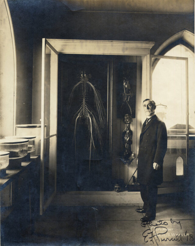
At Hahnemann, Weaver was appointed custodian of the university’s anatomical museum in 1880, and busied himself assembling an anatomical wunderkammer with no rival. Gone were papier-mâché models and “musty,” dried-out specimens. Weaver filled the light-flooded, third-floor space with hundreds of new medical displays, many of which he prepared himself. His trove included bladder calculi, sections of healthy and diseased brains, and an entire uterus, partly consumed by a tumor and opened to reveal a six-month-old fetus. The anatomist imagined these—and the museum’s hundreds of other objects—as teaching tools instead of “mere ‘curiosities,’” according to an announcement circulated in the mid-1880s. Among the assortment, there was Weaver, described in 1902 by a reporter from The North American as a “little professor” brimming with “energy, originality, and vim,” “as cheerful and bright as a May morning,” and prone to speaking of his collection of “beautiful tumor[s]” with tenderness and awe. (“Here is a lung,” the reporter quoted him saying. “Isn’t that the handsomest thing that you ever saw?”) In one 19th-century photograph, Weaver poses next to a fresh cadaver, its chest pried open, while limbs dangle around it like cuts of meat in a butcher’s shop. The anatomist’s own bearing was stick-straight—perhaps an occupational hazard of standing above so many spinal columns.
. . . .
But these guides stop well short of how Weaver pulled his masterpiece off. The earliest description of Weaver’s work on the nervous system comes courtesy of Thomas, who described the process in an 1889 edition of The Hahnemannian Monthly, the school’s journal. But Thomas’s is a hazy picture, long on the basics of dissection and short on clarity about how Weaver managed to preserve delicate nerve structures while chipping or sawing bone apart. This must have been finicky work: The spinal cord—a hardy nerve bundle—is roughly as wide as your thumb. We don’t have the complete ingredients Weaver mingled in his preservatives, a full inventory of the tools he enlisted, or a meticulous record of which parts of the process proved surprisingly straightforward or especially thorny or vexing. We don’t have a precise timeline, either. As Thomas tells it, dissection began on April 9 and concluded by June, with mounting complete by September; years later, van Baun reported that the dissection alone took nearly seven months, and then it required “seventy days of unceasing, laborious, skilled work and supreme patience to get the specimen on the board,” for a total of “nine months of gruelling [sic] contest.”
Weaver is said to have spent up to 10 hours a day in his humid office, and reportedly spent two weeks just tussling with the bottom of the skull. Once “all the little branching strands … were laid bare,” The North American noted, Weaver attempted to keep them supple by swaddling them in alcohol-soaked gauze or wads of cotton, which needed frequent changing, and he covered the flimsy strands with rubber. He retrieved nearly everything but sacrificed the intercostal nerves, which run along the ribs and proved too difficult to wrangle. Weaver reportedly excised the brain but held on to the outer membrane, called the dura mater, and plumped it up with “curled hair” stuffing, stitched it closed, and returned it to the display. To showcase the optic nerves, Weaver left the corpse’s eyes in place and distended them “with a hard injection,” Thomas wrote.
Mounting the specimen—as Weaver later recalled to The North American—was far more “wearisome and exacting” than the dissection itself. Weaver apparently tacked the nerves in place with 1,800 pins, and then fixed every filament with a coat of lead paint. (Many of those pins were later removed, Thomas wrote, once the shellacked nerves dried and held their position.) In all, Weaver reportedly spent several months laboring over the body, with a break for a summer vacation. The ultimate result, Thomas wrote, was “perfectly clean and free from all extraneous tissues and smooth as threads of silk.”
. . . .
Some laypeople argued that a living patient was better off being treated by someone who had seen the body’s inner contents up close. In 1882, The Christian Recorder—the newspaper of the African Methodist Episcopal Church—endorsed dissection, suggesting that it would be foolish for anyone to seek treatment “at the hands of a man who had not gone through the mysteries of the dissecting room.” Still, even those who supported the notion of dissection typically did not want to entertain the thought of it happening to anyone they loved. The anonymous author of that article in The Christian Recorder skewered grave robbing on moral grounds and suggested that doctors be offered the bodies of executed murderers and anyone who died by suicide.
The few people who expressly permitted, or even beseeched, doctors to cut into them after death tended overwhelmingly to be white, wealthy, and accomplished men. By 1889, the new American Anthropometric Society, headquartered in Philadelphia, began compiling the brains of physicians and public intellectuals who embraced the ideas of phrenology, which correlated intellectual feats with cranial attributes. These donors were keen to join the organization’s “brain club” as a way to further the field while also valorizing themselves.
And in the medical realm, consent was slippery. William Osler, a founding professor of Johns Hopkins Hospital, was known to solicit family approval before giving cadavers to his students—but he was also famously dogged in his pursuit of that permission, and in a 2018 article in the journal Clinical Anatomy, Wright, the University of Calgary pathologist, notes that “autopsy consent and organ retention abuse was not uncommon in late-19th century Philadelphia.” In a 2007 Academic Medicine article about the uptick in body bequeathal in 20th-century America, Ann Garment, then a medical student at New York University, and three coauthors note that turn-of-the-century body donation was uncommon enough to make the news when it happened. The New York Times picked up the tale of Thomas Orne, a wealthy Maryland horse dealer who pledged his body to Johns Hopkins in 1899. In 1912, 200 New York City physicians also vowed to donate their bodies for dissection in an effort to erode the stigma around it.
. . . .
“I am aware that there have been men, [the philosopher Jeremy] Bentham for instance, who have voluntarily willed their bodies to be dissected, but they have been extremely few,” Sozinsky recounted in 1879. Opting in was far from commonplace. “The ‘Harriet Cole’ story, if correct, is likely very unusual,” Wright notes. If a flesh-and-blood Black woman named Harriet Cole consented to her own dissection more than 130 years ago, she would have had very little company.
Link to the rest at Atlas Obscura